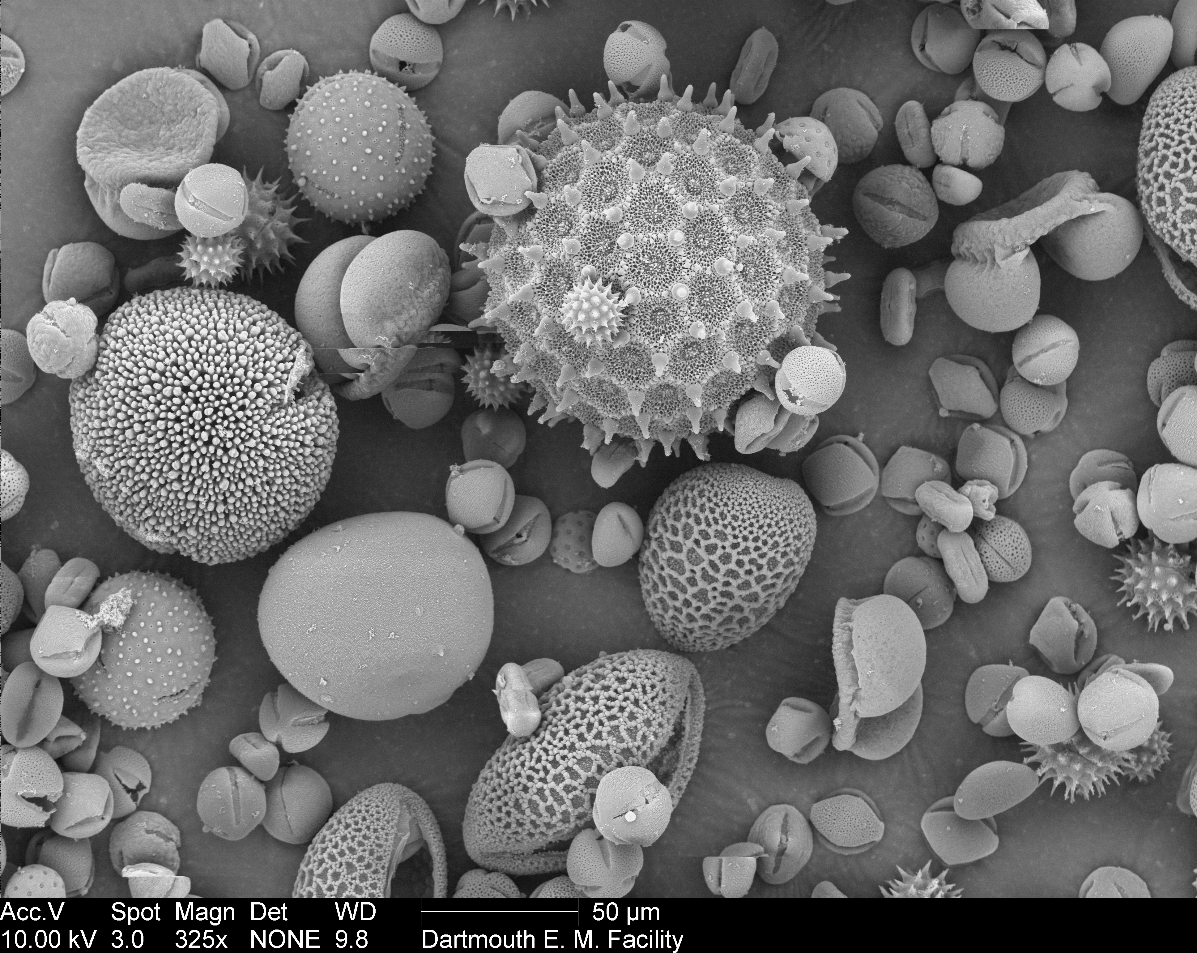
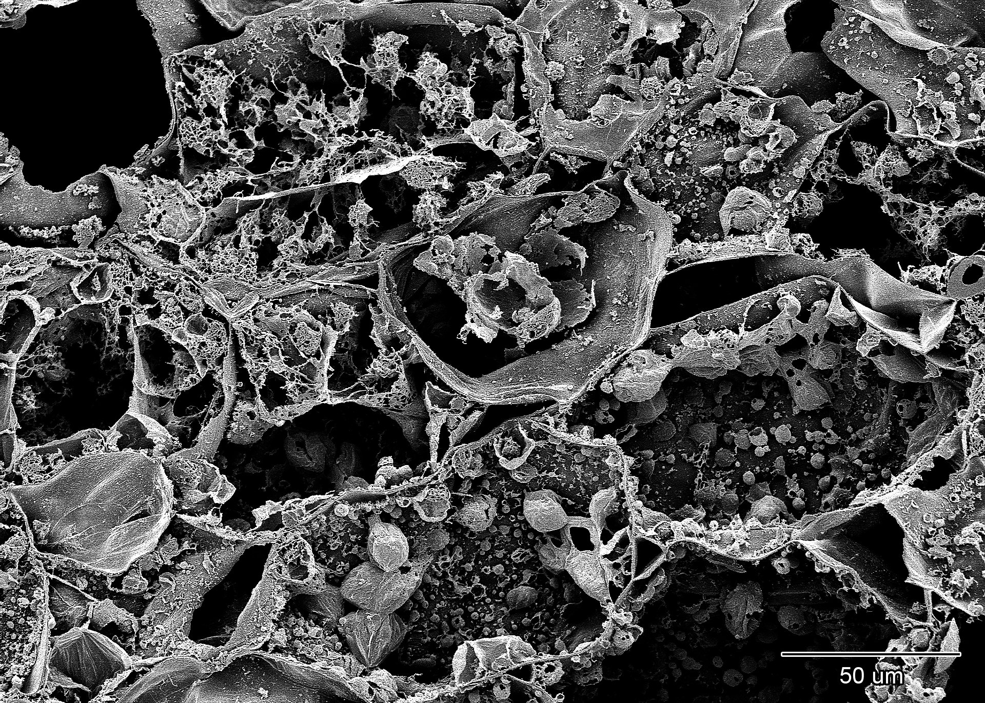
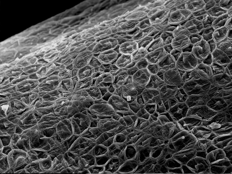
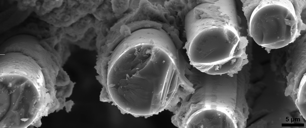
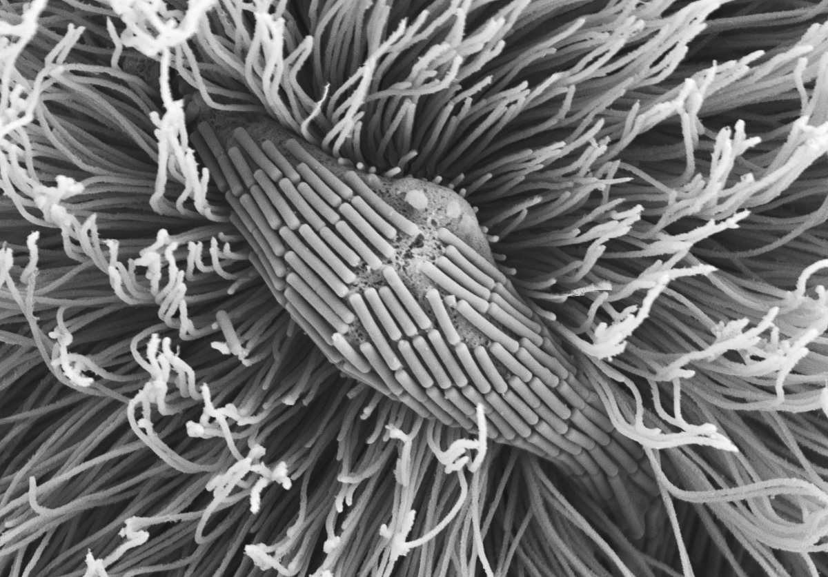
Как выглядят микроспоры под электронным микроскопом - смотри 30 фото
Найдено фото: 30
Thu., Sep. 5 notesКартинки МИКРОСКОПИЯ ТЕЛА
Чернозем под микроскопом - фото и картинки abrakadabra.funКристаллы Льда под Микроскопом. Часть 2. Электронный микроскоп / XpathHome Bioscience Electron Microscopy LaboratoryPeering into the micro world Electron microscope images, Scanning electron microBiochar for Commercial Horticulture - Carbon Gold Tree care, Carbon sequestratioVirus under Scanning Electron Microscope
The complexity and intricacy of Mother Nature revealed by incredible pictures ofFantastic Voyage Electron Micrograph For the Home Photography, Micro photographyМикромир (фото) Electron microscope, Electron microscope images, Microscopic imaA peculiar association between a coccolithophorid haptophyte (small phytoplanktoFIG URE 8 Scanning electron microscope ... Scanning electron microscope, ElectroSingle-Celled Eukaryote Fossil with Mineralizing Evidence in Yukonelectron microscopy alveoles count Braided rugs, Things under a microscope, Home
root tip cell Electron microscope, Plant cell, Electron microscope imagesTricomes on Squash leaf surface_2 Scanning electron microscope, Electron microscFlower - electron microscope Turning away from the big, the grand, the monumentaA marine diatom, surrounded by coccoliths. Macro and micro, Diatom, OceanographyPeering into the micro world Scanning electron micrograph, Micro photography, PeMore bacteria are becoming resistant to antibiotics - here's how viruses and vacWarm waters melting Antarctic ice shelves may have appeared for the first time iDuke Neurology Research Round Up, February 2019 - Duke Center for NeurodegeneratСегодня многие начинают задумываться над тем, что в партнерстве и дипломатии конEye of the greenfly - University technologist scoops award for striking image NeTransmission Electron Microscope Service J.Kraft MicroscopyThe ocean under a microscope: Carla Stehr merges art and science South Whidbey RInvisible Portraits