Микропрепарат растительной клетки под микроскопом - смотри 30 фото
Найдено фото: 30
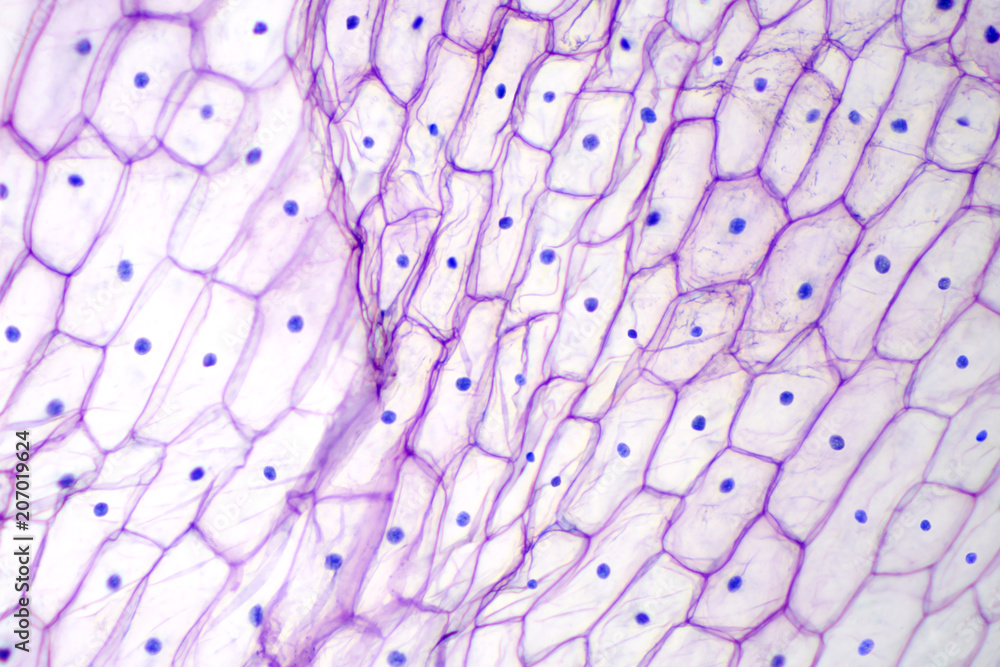
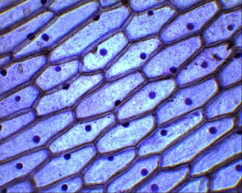
ГДЗ биология 5 класс учебник с шишкой ПасечникOnion epidermis under light microscope. Purple colored, large epidermal cells of
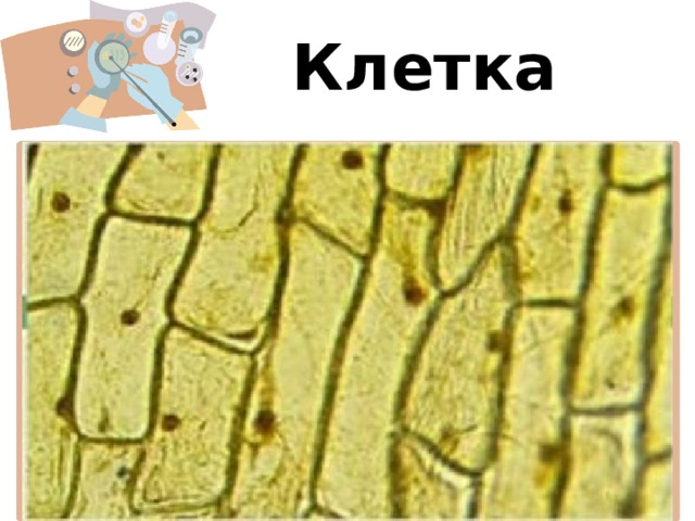
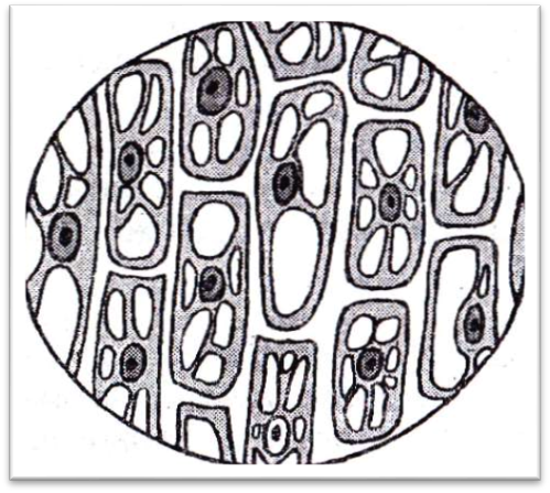
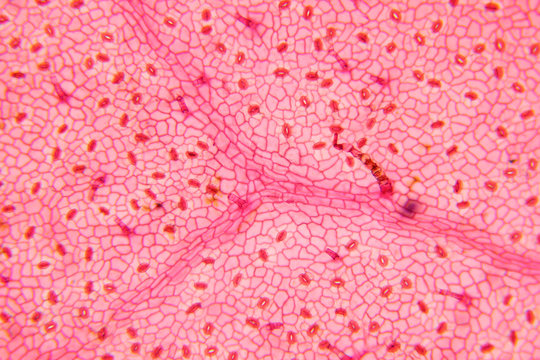
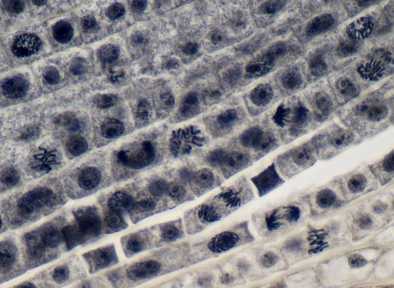
Микроскоп "Юный исследователь", увеличение х1200 - купить в интернет-магазине попрезентация строение клетки 5 классКлетка тирүү организмдердин түзүлүшү жана тиричилик бирдигиLongitudinal section of stained plant cell stomata Natura, Biologia, ScienzaMitosis, Science art, Science jokesOnion epidermis with large cells under light microscope. Clear epidermal cells o
plant cells under a microscope - Google Search Biology art, Art, Plant cell35 Stunning Examples Of Photomicrographs Mitosis, Microscopic photography, PatteTop Tips for Observing Mitosis Lab Mitosis, Meiosis, Meiosis activityLearn About Plant Cell Types and How They're Like Animal Cells Plant cell, MicroValuation under the microscope Microscopic photography, Microscopic images, ThinPin by tua on textures Microscopic photography, Cells worksheet, Teaching sciencBio Blogger: Microscope Lab
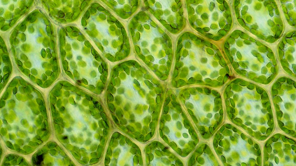
Gene circuits in live cells can perform complex analogue and digital computationSpecialised plant cells - Living organisms - KS3 Biology - BBC BitesizeМикроскопия картинки - 30 фотоТкани растений - строение и функции в таблице, виды и типыДеление клетки (митоз) на примере препарата кончика корешка лука - "Sky-Route"Miracles of Biophotonics "Tomorrow" and "Tomorrow". Sergey Vladimirovich, now ouCells Under Microscope Зображення - огляд 31,254 Стокові фото, векторні зображенФайл:Root tip.JPG - ВикипедияФайл:Meristemo apical 1.jpg - ВікіпедіяГалерея фотографий. Что можно увидеть в микроскоп Levenhuk 630Наборы готовых микропрепаратов Levenhuk442 Red Onion Cell Images, Stock Photos & Vectors Shutterstock15,865 Plants Dna Images, Stock Photos & Vectors ShutterstockКлетки и ткани