Микропрепарат животной клетки под микроскопом - смотри 30 фото
Найдено фото: 30
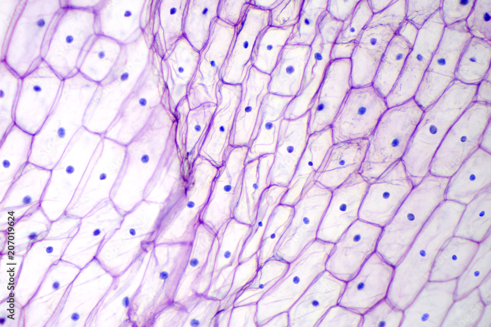
Onion epidermis under light microscope. Purple colored, large epidermal cells ofМикроскоп "Юный исследователь", увеличение х1200 - купить в интернет-магазине по
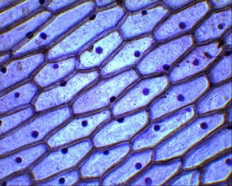
Pin by Curly Tea on Nurse Microscopic, Cardiac muscle cell, MuscleMitosis, Science art, Science jokesOnion epidermis with large cells under light microscope. Clear epidermal cells oSimple Squamous Epithelium, flat mount Tissue, Tissue types, Nursing tips35 Stunning Examples Of Photomicrographs Mitosis, Microscopic photography, PattePin on microscopico
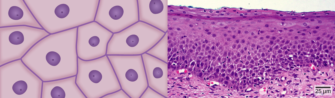
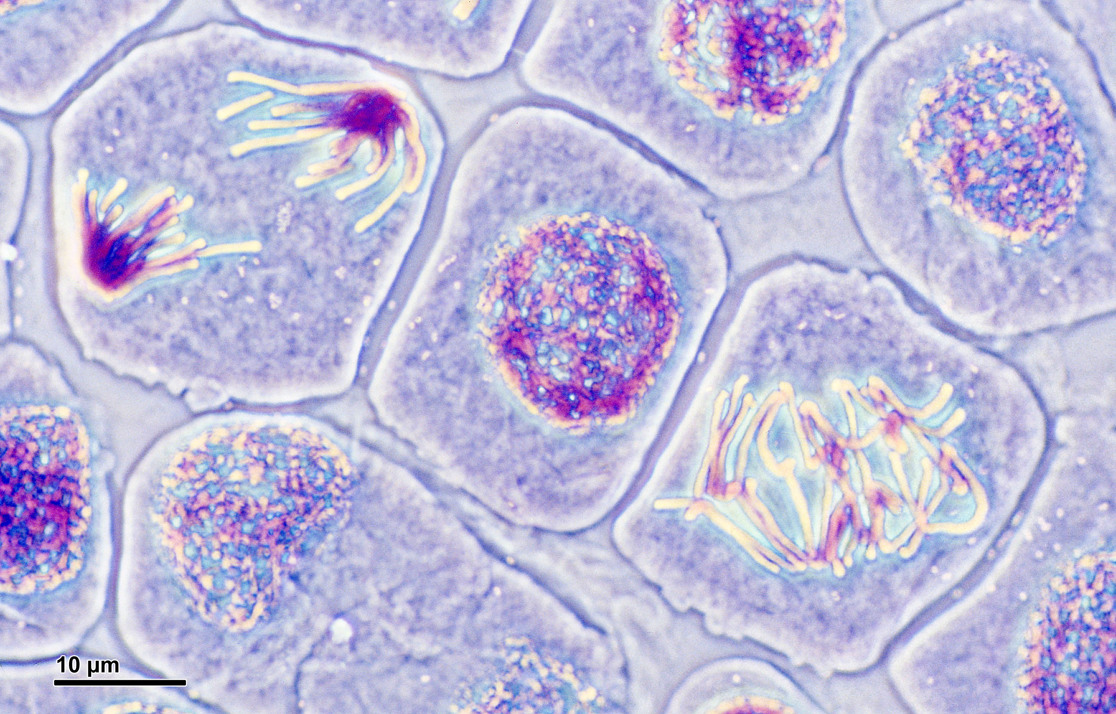
Top Tips for Observing Mitosis Lab Mitosis, Meiosis, Meiosis activityГистологические окраски 2 Anatomy art, Scientific illustration, Medical school sValuation under the microscope Microscopic photography, Microscopic images, ThinMitosis, Teaching biology, Biology artPin by tua on textures Microscopic photography, Cells worksheet, Teaching sciencЭпителиальная ткань под микроскопом рисунокBio Blogger: Microscope Lab
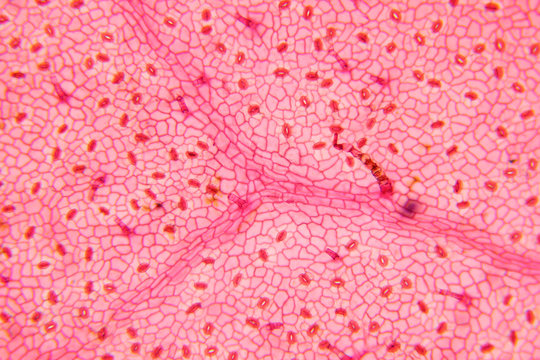
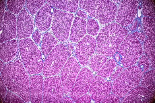
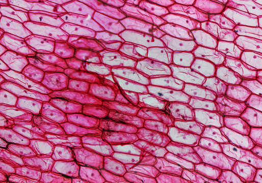
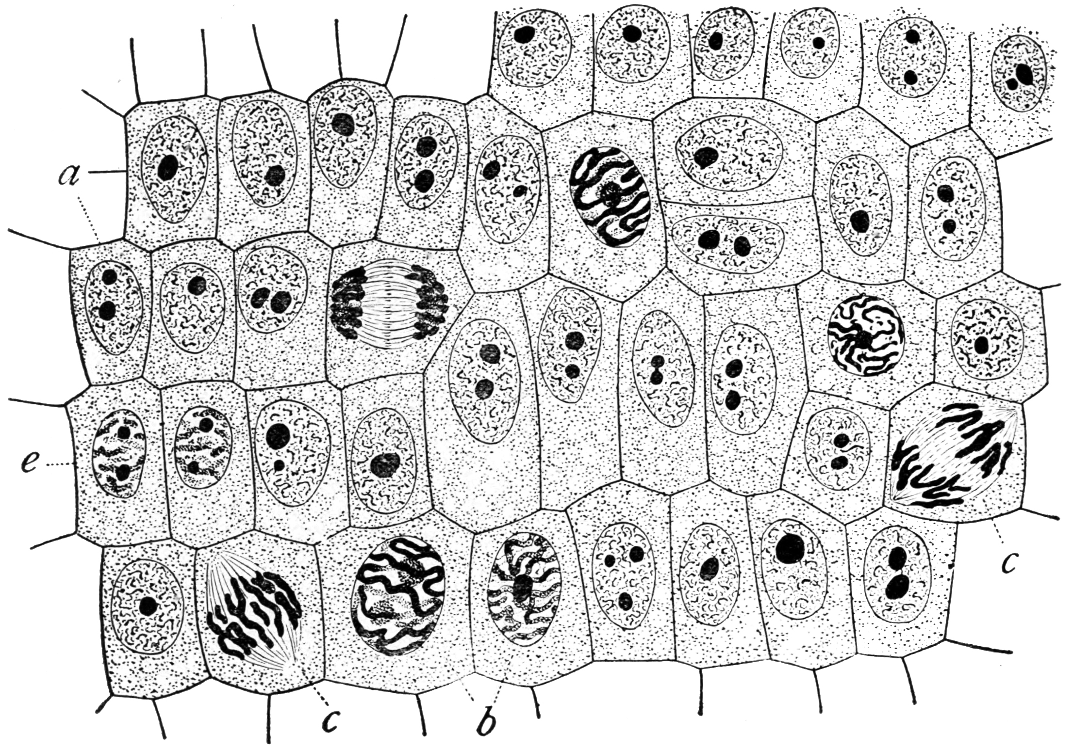
microscopic images of plant and animal cells - Google Search Plant and animal ceGene circuits in live cells can perform complex analogue and digital computationМикроскопия картинки - 30 фотоCell Biology 1 Flashcards QuizletТкани растений - строение и функции в таблице, виды и типы14.2 Animal Primary Tissues - Concepts of Biology-1st Canadian Edition Molnar Clцитология - 2 фотографии ВКонтактеCells Under Microscope Зображення - огляд 31,254 Стокові фото, векторні зображенMicroscope Histology Зображення - огляд 21,067 Стокові фото, векторні зображенняSkin Cells Изображения: просматривайте стоковые фотографии, векторные изображениFile:Mitosis (261 11) Pressed; root meristem of Vicia faba (cells in anaphase, pФайл:Wilson1900Fig2.jpg - ВикипедияFile:Onion Cells.jpg - Wikipedia442 Red Onion Cell Images, Stock Photos & Vectors ShutterstockКлетки и ткани