Микроскоп "Юный исследователь", увеличение х1200 - купить в интернет-магазине поКнижно-иллюстративная выставка "Клеточный мир" 2019, Пошехонский район - дата и Stunning Microscopic View of Human Skin Cells Wins 2017 Nikon Small World CompetМир под микроскопом
Строение животной клетки.Новое микрофото (con imágenes)Learn About 11 Different Cell Types in the Body Body cells, Cell, BodyOnion epidermis with large cells under light microscope. Clear epidermal cells oBBC - Earth - The secret of how life on Earth began Life on earth, Earth, SciencWelcome to the IPBio Network Microscopic photography, Microscopic images, ThingsPin by Sarah Bell Smith on collections of images Microscopic photography, Micros
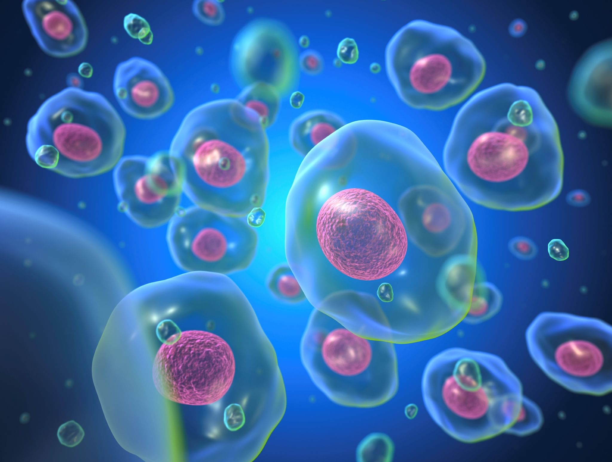
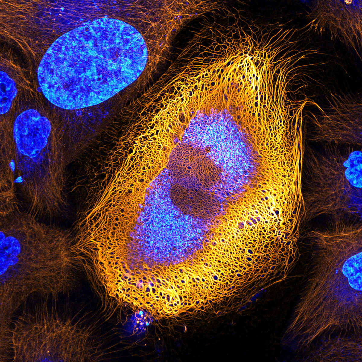
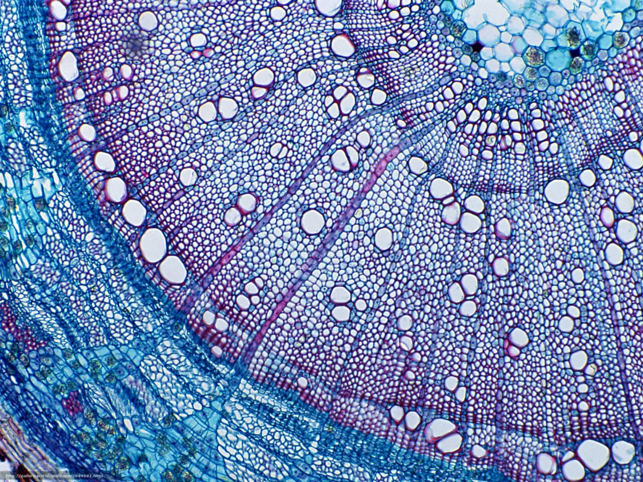
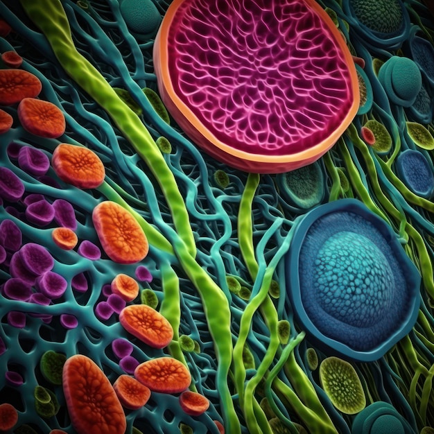
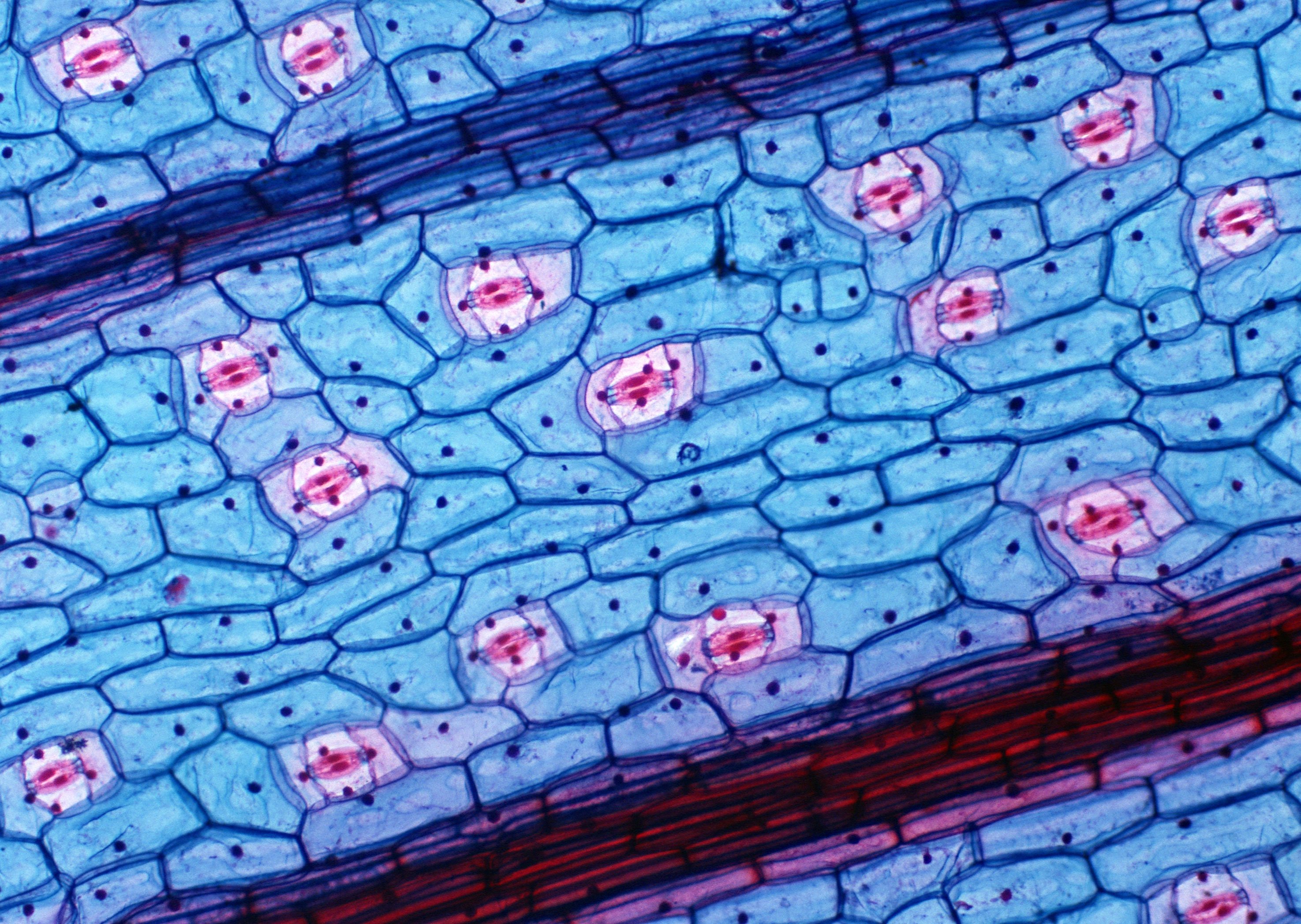
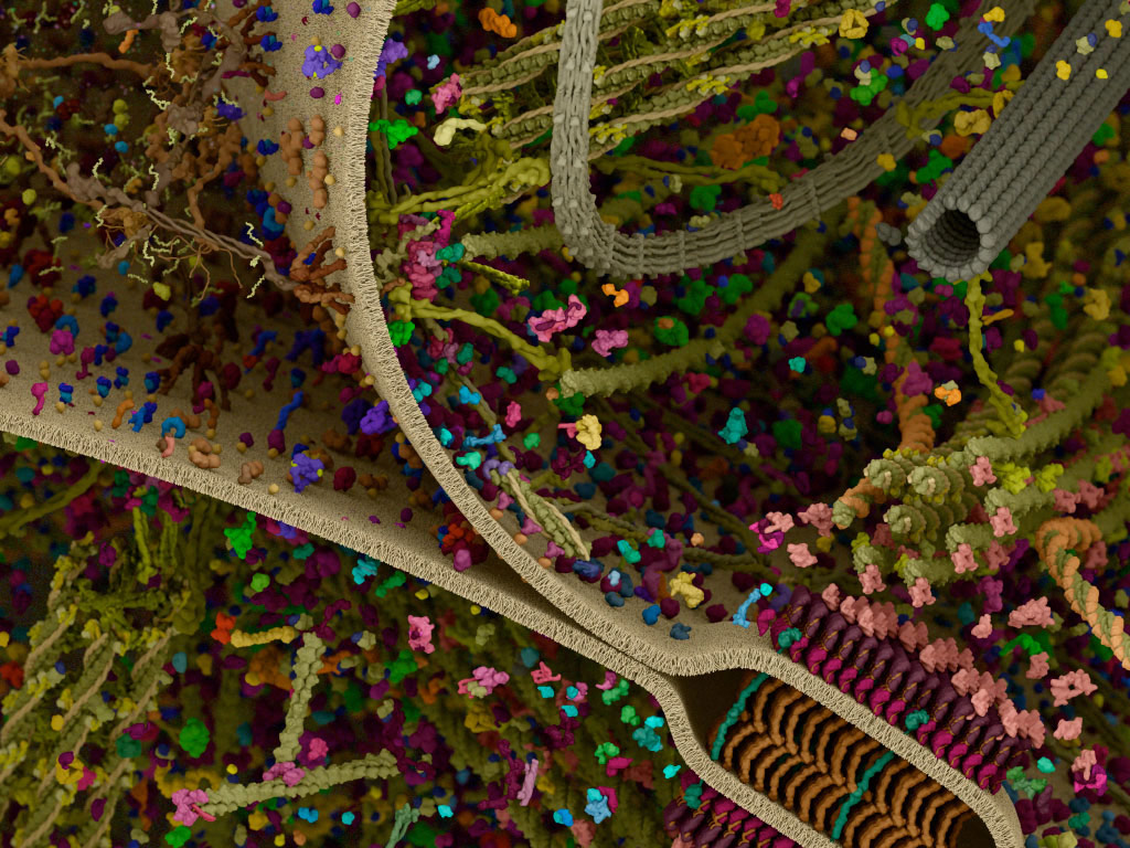
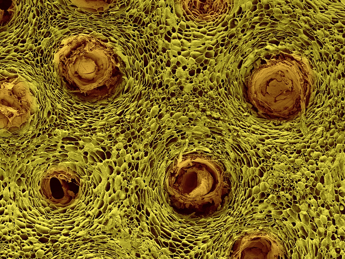
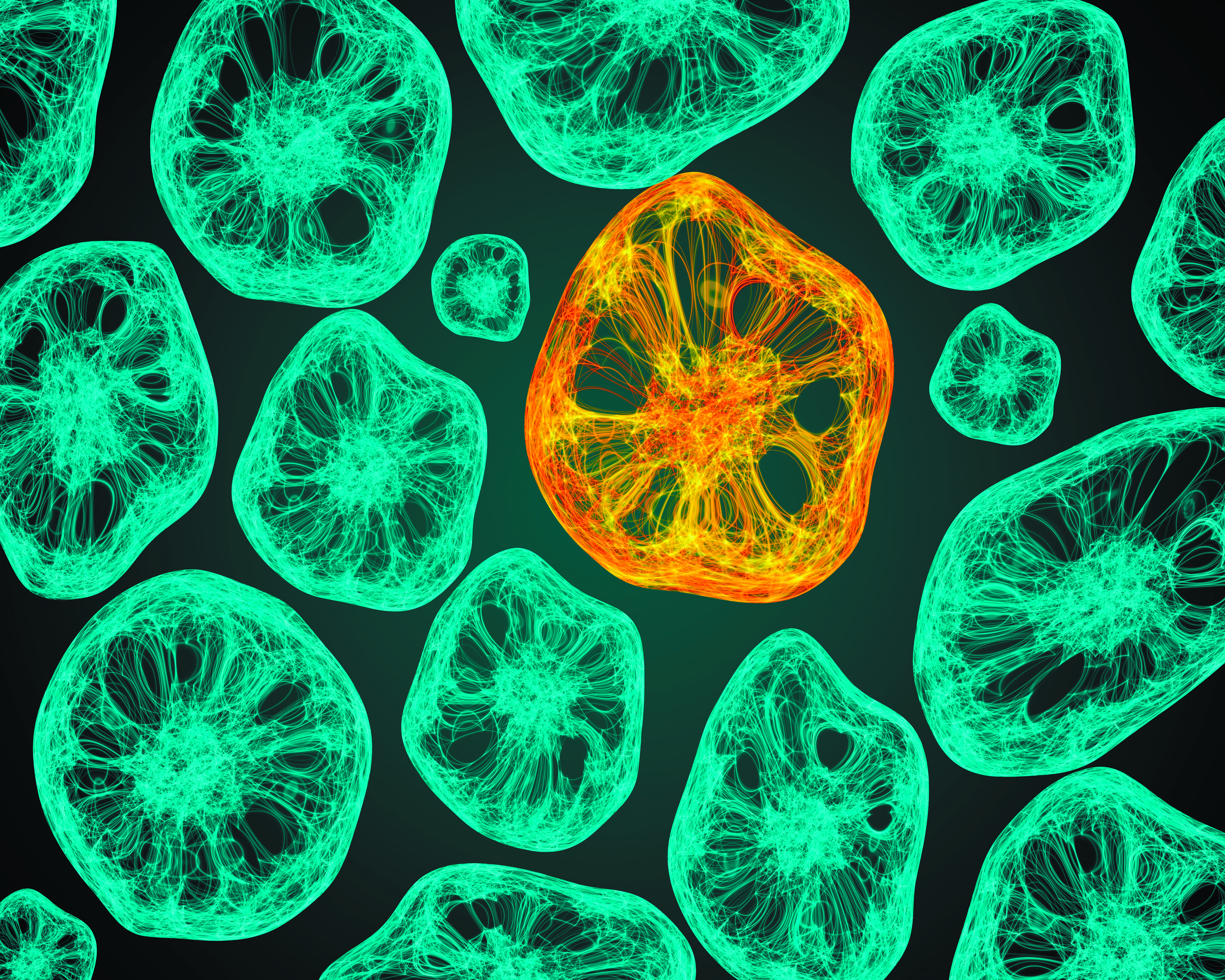
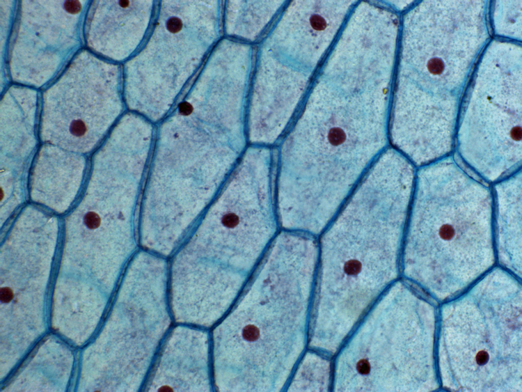
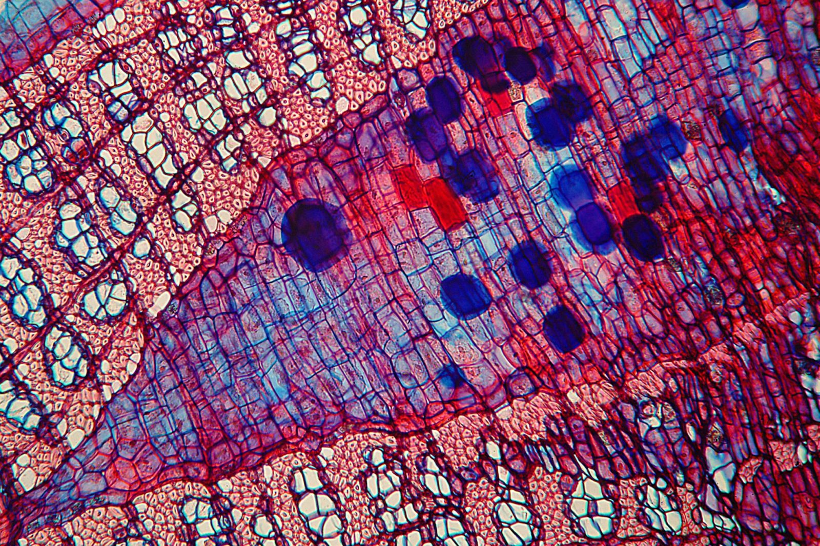
Animal Cells Educational Resources K12 Learning Animal cell, Animal cell structuPoster Print Wall Art Print entitled Cell Poster prints, Wall art prints, Art pr17 Mind-Bending Pictures Of Life Through A Microscope Human brain, NeuroplasticiCellular Landscapes: Protein Synthesis Cell Signaling Technology in 2024 StructuPin on Guardado rápidoThings under a microscope, Shape and form, Microscopic imagesEpidermis, Lcd, PixelSpectacular Microscopic Art Is Also World-Changing Science Microscopic photograpNikon MicroscopyU Small World Competition 1985 Microscopic cells, Nikon small woFull HD. Many living dividing cells under microscope, magnification 400X Stock FPlant Cell Microscopy http://www.flickr.com/photos/anhedonias/ Microscopic photoValuation under the microscope in 2022 Microscopic photography, Biology art, MicNeuronally differentiated P19 cells 2009 Photomicrography Competition Nikon smalCells artunderwater coral photography - Google Search Marine life, Sea and ocean, Time laAnd these are our lung cells. Biology art, Things under a microscope, MicroscopiCross Section of the stem of a baby pine tree. Patterns in nature, Fractal art, Cross Section of the stem of a baby pine tree. Patterns in nature, Fractal art, Here cultured epithelial cells are imaged via multiphoton microscopy, illustratiМикроскопия картинки - 30 фотоMolecular Expressions Microscopy Primer: Specialized Microscopy Techniques - FluВ МФТИ изучили внутриклеточную передачу сигнала при помощи разделения фазУглубленное изучение биологииТкани растений - строение и функции в таблице, виды и типыКартинки РАСТИТЕЛЬНАЯ КЛЕТКА СВЕТОВОЙ МИКРОСКОПΚατηγορία:Κύτταρα Science Wiki FandomΚατηγορία:Κύτταρα Science Wiki FandomПубликация #335 - I (PLlmYSxVsTM2NjMy)Периферический нерв: pigmentum ВКонтактеВидео кожицу: найдено 75 изображенийCells Under Microscope Зображення - огляд 31,254 Стокові фото, векторні зображенПлатформа трансфера технологий ИТМОFile:Onion Cells.jpg - WikipediaТкани растительного организма под микроскопомBiology CUNY Graduate CenterВ ИТМО разработали сервис для анализа биохимических реакций в клетках на уровне Appie Bonis People Nikon’s Small WorldCell Wall Structure and FunctionCustom-tailored strategy against glioblastomas - University of BonnCollection of 10 Microscope Preparations Botany Cytology


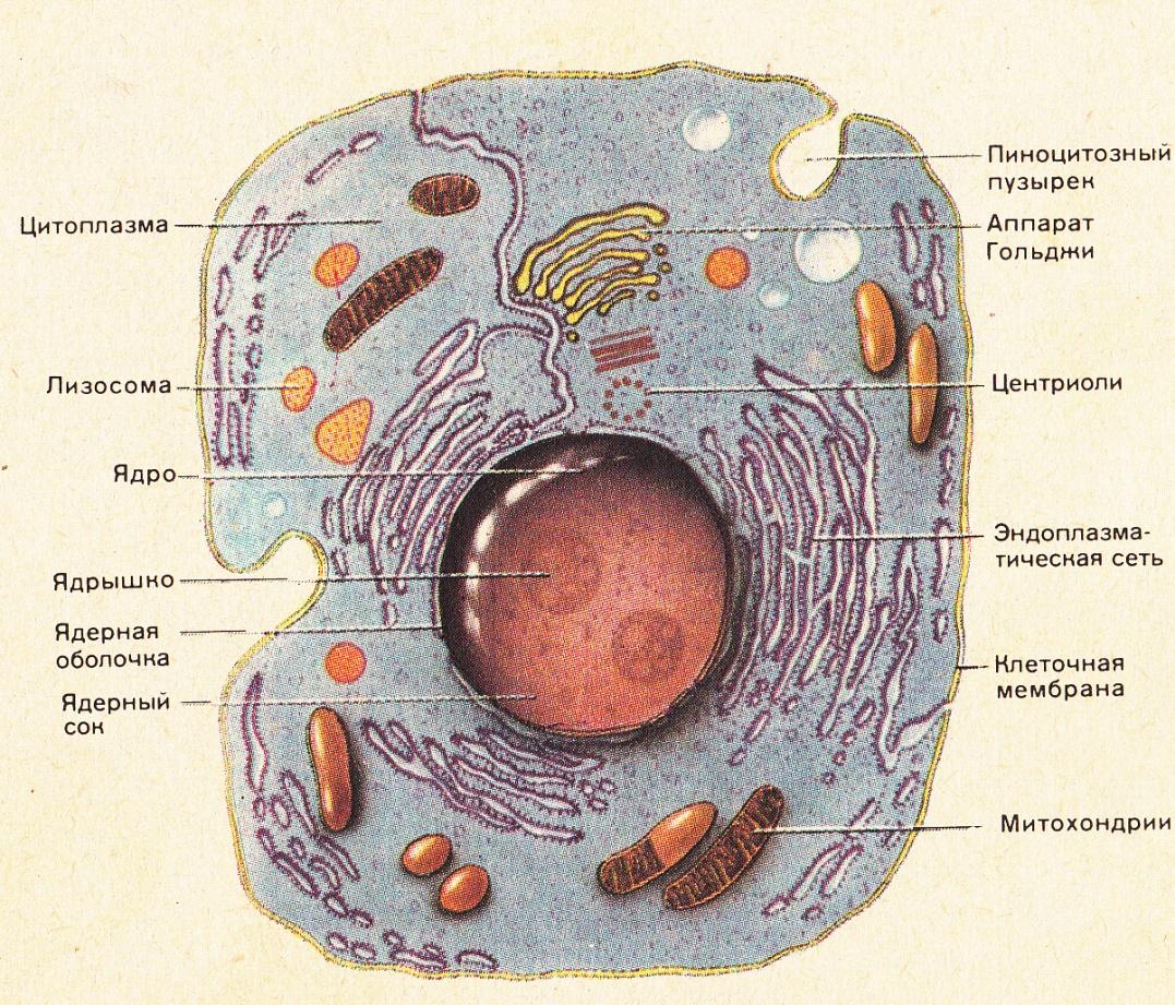


:max_bytes(150000):strip_icc()/plant_cell_organelles-5b64a69f46e0fb00253a8bf4.jpg)